
World Pneumonia Day 2026: What Lung Imaging Reveals About Pneumonia
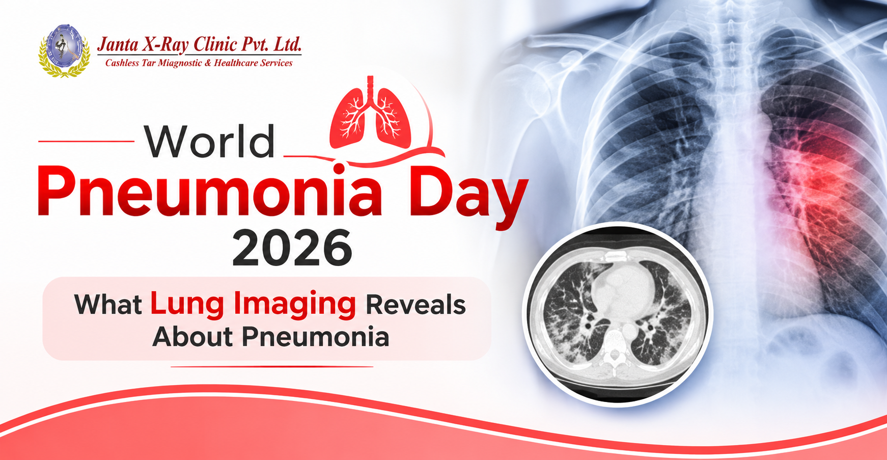
Pneumonia is not simply a respiratory infection that makes breathing uncomfortable. It affects the very structures responsible for oxygen exchange within the lungs. When infection causes inflammation in the tiny air sacs, known as alveoli, these spaces may fill with fluid, making normal breathing increasingly difficult.
Observed on 12 November, World Pneumonia Day highlights the importance of recognising this condition and understanding how timely medical evaluation can help prevent complications. Persistent cough, fever, chest discomfort and breathlessness are common symptoms, but their presence alone cannot establish whether someone has pneumonia.
What Changes Inside the Lungs During Pneumonia?
Healthy lungs depend on millions of air sacs functioning efficiently. During pneumonia, inflammation disrupts this process, affecting the movement of oxygen into the bloodstream. Depending on the infection, the affected area may be limited to one section of a lung or extend across multiple regions. These changes are important because the location and extent of lung involvement can influence clinical assessment and treatment decisions.
What Can a Chest X-Ray Reveal?
A Chest X-Ray for pneumonia is commonly recommended when symptoms and clinical examination raise suspicion of a lung infection. It helps identify abnormalities such as consolidation, where normally air-filled lung tissue appears denser because of fluid or inflammatory material. Radiologists also examine the distribution of these changes and look for associated findings, including fluid around the lungs.
However, interpreting a Chest X-Ray involves more than identifying white patches on an image. Their location, appearance and distribution must be considered alongside the patient's clinical condition. An early infection may also not be clearly visible on an initial X-Ray.
When Does CT Chest Become Important?
When a Chest X-Ray does not adequately explain persistent symptoms, or when complications are suspected, a doctor may recommend CT Chest. This investigation provides detailed cross-sectional images that help assess lung tissue, the extent of abnormalities and certain complications. It is not routinely required for every pneumonia patient; its use depends on clinical necessity.
Why Radiological Interpretation Matters
Pneumonia assessment is not just about identifying an abnormality. It is about understanding what that abnormality could indicate and how it relates to the patient's symptoms. At Janta X-Ray Clinic Pvt. Ltd., radiological imaging supports the evaluation of chest and lung conditions through relevant imaging investigations and professional interpretation.
On World Pneumonia Day, it is worth remembering that persistent respiratory symptoms deserve medical attention. The right imaging investigation, recommended at the appropriate time, can provide valuable information about lung health and support doctors in making informed clinical decisions.
For doctor-recommended chest imaging investigations, contact Janta X-Ray Clinic Pvt. Ltd. at 011-49114911 or visit jantaxray.in
Frequently Asked Questions
Question: Can the location of an abnormality on a Chest X-Ray help assess pneumonia?
Answer: Yes. The distribution of lung opacities can help radiologists assess which regions are affected and provide useful information for clinical evaluation.
Question: Why might a patient with pneumonia need another Chest X-Ray?
Answer: A doctor may recommend follow-up imaging in selected cases to assess changes in lung abnormalities, particularly when symptoms persist or recovery needs further evaluation.
Question: Can a Chest X-Ray distinguish bacterial pneumonia from viral pneumonia?
Answer: No. Although certain imaging patterns may offer clues, a Chest X-Ray cannot reliably identify the organism responsible. Clinical assessment and appropriate laboratory investigations are needed.
Question: Does every abnormal lung opacity indicate pneumonia?
Answer: No. Lung opacities can occur due to several conditions, including infection, inflammation and fluid accumulation. Their interpretation requires clinical context and radiological expertise.










Comments List