
When the Diagnosis Is Hidden Between the Lines
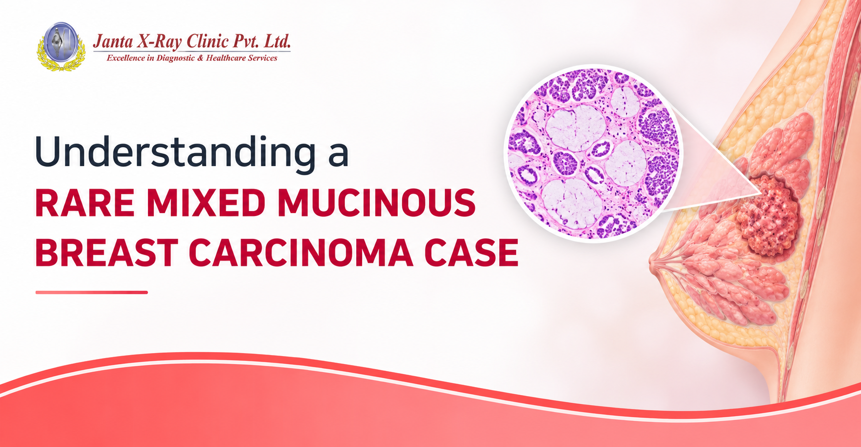
For patients and families: A breast lump can require more than one test before doctors and pathologists can understand exactly what it is. This case shows how FNAC, core biopsy, surgery, histopathology and immunohistochemistry can each contribute a different piece of the diagnosis.
The key message
An inconclusive FNAC does not by itself rule out breast cancer. When clinical or imaging findings remain concerning, further tissue evaluation may be needed.
A rare diagnosis, explained simply
Mucinous carcinoma is an uncommon type of breast carcinoma, also described as colloid, mucoid or gelatinous carcinoma. It can occur in a pure form or together with an invasive ductal component. The supplied case report describes pure mucinous carcinoma as having at least 90% mucinous component. When mucinous and invasive ductal areas occur together, the tumour is described as a mixed form.
What happened in this case?
A 35-year-old woman presented with lumps in both breasts that had been present for four months. There was no family history of breast cancer. On examination, the left breast had a firm, tender 4 × 4 cm lump that was fixed to the overlying skin. The right breast had a firm, freely mobile 5 × 5 cm lump.
Why the first test did not give the whole answer
Fine-needle aspiration cytology (FNAC) examines cells obtained with a thin needle. In this case, FNAC of the left breast was reported as carcinoma breast (C5). FNAC of the right breast was inconclusive on two occasions.
Importantly, the cytology showed ductal epithelial cells dispersed within pools of extracellular mucin. This was an important clue pointing toward a mucinous tumour. However, FNAC may not always show enough of the tissue architecture needed to fully characterize a breast lesion.

Figure 1. Cytosmear showing ductal epithelial cells dispersed in pools of extracellular mucin (H&E, 4×).
Core biopsy added another piece of the puzzle
Core-cut biopsy was performed from both breasts. The left breast was suggestive of mucinous carcinoma, while the right breast showed occasional benign breast ducts in fibrocollagenous stroma.
The patient subsequently underwent left modified radical mastectomy with axillary clearance and lumpectomy with wide local excision of the right breast.
What the surgical pathology showed
The left mastectomy specimen contained firm-to-hard areas as well as a separate soft, well-defined area. Under the microscope, tumour cells were arranged in nests and cords and were also present within pools of mucin, forming clusters and acini. The cells showed pleomorphism and hyperchromatic nuclei, with occasional mitotic figures.
The case was characterized as a mixed mucinous carcinoma because mucinous areas were seen together with an invasive ductal component. The final histopathological diagnosis recorded in the supplied report was invasive ductal carcinoma with mucinous differentiation.

Figure 2. Mixed mucinous carcinoma comprising invasive ductal and mucinous components (H&E, 4×).

Figure 3. Tumour cells floating in pools of mucin along with invasive ductal carcinoma (H&E, 10×).
What happened to the lymph nodes and margins?
The deep and peripheral surgical margins were free of tumour. Twenty-one lymph nodes were identified and all were free of tumour.
Why were ER, PR and HER2 tested?
Immunohistochemistry showed negative estrogen receptor (ER), progesterone receptor (PR) and HER2/neu status. These tests provide additional information about the biological characteristics of a breast tumour and are used to support clinical management planning.

Figure 4. Mucicarmine positivity in mucinous areas.
And what about the lump in the right breast?
The right breast lumpectomy specimen contained two nodules measuring 6 × 4.5 × 4 cm and 5 × 3 × 1.5 cm. Microscopy showed benign breast ducts and acini within dense fibrocollagenous stroma. Some ducts were cystically dilated with eosinophilic secretions, with focal adenosis and epithelial hyperplasia.
The supplied case report states that the patient remained under regular follow-up and had received two cycles of chemotherapy.
What does this mean for patients?
- A breast lump should be evaluated appropriately rather than judged by one test alone.
- FNAC can provide an early cytological clue, including the presence of extracellular mucin.
- If FNAC is inconclusive, further tissue evaluation such as core biopsy may be needed.
- Histopathology helps doctors see the overall tissue architecture and the relationship between different tumour components.
- ER, PR and HER2 testing adds biological information that can help clinicians plan management.
- A diagnosis is built by putting the clinical findings and different laboratory and pathology results together.
Frequently asked questions
Can FNAC diagnose mucinous breast carcinoma?
FNAC can provide an important clue. In this case, ductal epithelial cells were seen within pools of extracellular mucin. However, core biopsy and surgical histopathology helped establish the complete diagnosis.
What does extracellular mucin mean on a breast FNAC?
Extracellular mucin can be an important clue toward mucinous carcinoma. It needs to be interpreted together with the cellular features and the other clinical and pathological findings.
Why can FNAC sometimes be inconclusive?
FNAC samples cells rather than a larger piece of tissue. It may therefore not provide enough information about tissue architecture. When the findings remain inconclusive, a core biopsy or another appropriate tissue evaluation may be recommended.
What is mixed mucinous carcinoma of the breast?
It refers to a tumour containing both mucinous and invasive ductal components. Careful histopathological assessment is needed to identify how these components are related.
Why are ER, PR and HER2 tested?
These markers are assessed by immunohistochemistry to provide additional information about the biological characteristics of a breast tumour and to support clinical management planning.
Does an inconclusive FNAC mean there is no cancer?
No. An inconclusive FNAC does not by itself exclude breast malignancy. If clinical, imaging or other findings remain concerning, further evaluation may be required.
What this case teaches us
The most important lesson is that pathology is a process of correlation. Cytology may provide the first clue, core biopsy can add tissue information, surgical histopathology can demonstrate the tumour architecture, and immunohistochemistry can add biological characterization. Together, these findings can provide a more complete understanding of a breast lesion.

Dr. Priyanka Anand, HOD Lab and Senior Pathologist at Janta X Ray Clinic Pvt. Ltd., brings more than 11 years of experience in pathology. This case reflects an important principle of diagnostic medicine.
For anyone with a breast lump
Do not try to determine the nature of a breast lump from symptoms or a single test result alone. Your clinician will decide which combination of examination, imaging and tissue tests is appropriate for your situation.


.png)
.png)


.jpg)

.jpg)
.jpg)
Comments List