
How to Read a PET-CT Report: SUV, FDG Uptake and More
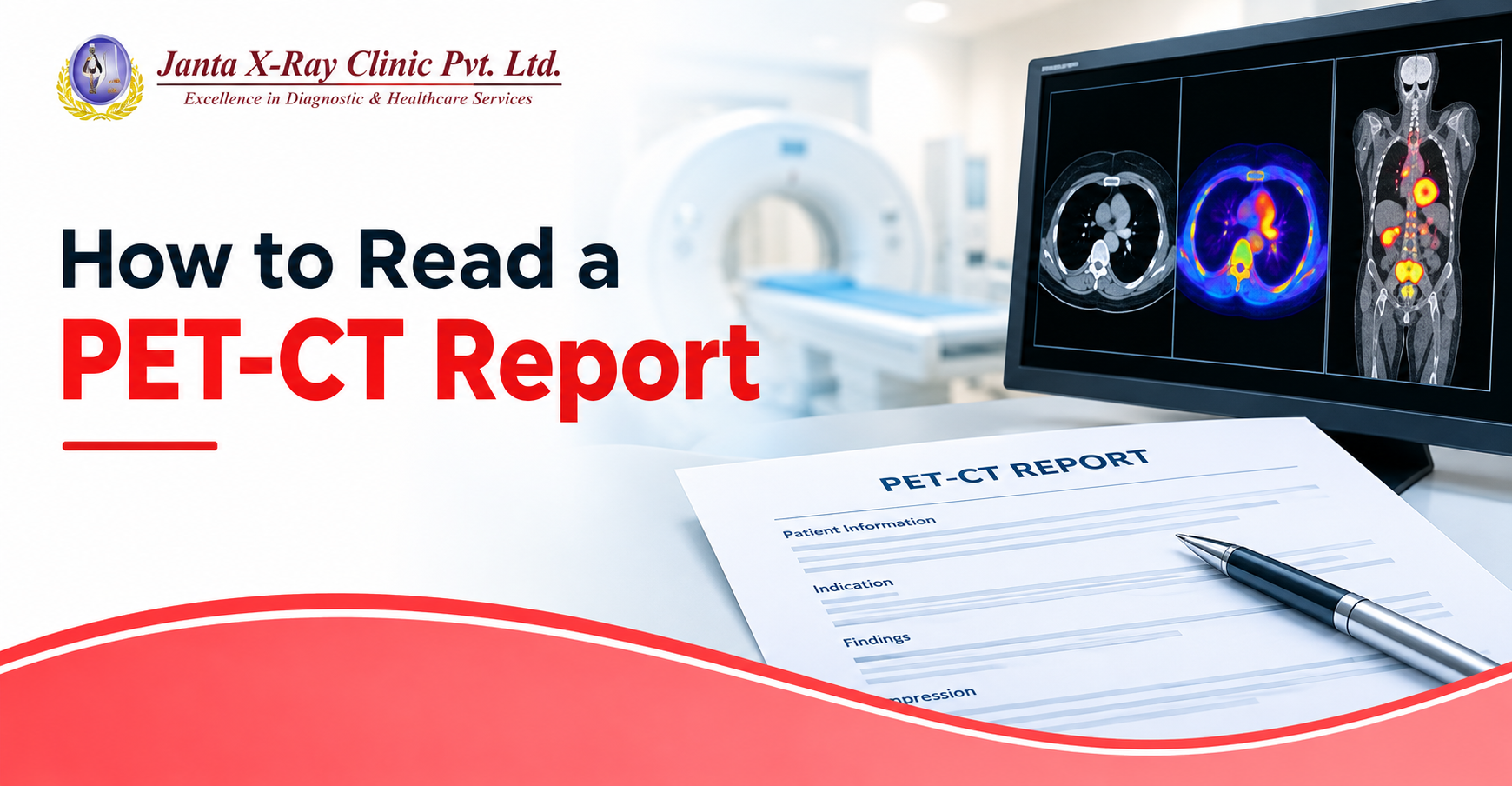
After you receive your PET-CT scan report you might find words that're hard to understand at first. Words such as FDG uptake, SUVmax, activity, FDG avid lesion, lymphadenopathy and metastatic disease can make the PET-CT report feel more complicated than it actually is.
That does not mean that every word that looks abnormal is cancer. The PET-CT report combines information, from PET and CT images. The findings must be considered together with your medical history and other tests.
Use this PET-CT report checklist to understand the terms you may see
PET-CT Report Checklist: What Does Each Term Mean?
☐ 1. FDG
FDG stands for fluorodeoxyglucose. It is a glucose-like radioactive tracer commonly used for PET scans.
After it is given, the tracer travels through the body and collects in tissues that use glucose. PET imaging then shows where the tracer has accumulated.
In simple words: If you see FDG in your PET-CT report, it is referring to the tracer used to show metabolic activity in the body.
☐ 2. FDG Uptake
FDG uptake tells you how much of the tracer has collected in a particular area.
A report may describe uptake as mild, moderate or intense. It may also mention increased FDG uptake in a particular lesion, lymph node or organ.
Increased uptake does not automatically mean cancer. Infection, inflammation and some normally active tissues can also take up FDG.
In simple words: FDG uptake shows where the tracer is collecting and how active that area appears on the PET scan.
☐ 3. Physiological FDG Uptake
You may come across the phrase physiological uptake in a PET-CT report.
This generally refers to FDG activity that is expected as part of normal body function. The brain, heart, kidneys, urinary tract and some other tissues can normally show FDG uptake.
In simple words: Physiological uptake usually means the activity is considered normal for that particular part of the body.
☐ 4. SUV
One of the most commonly noticed numbers in a PET-CT report is SUV, which stands for Standardized Uptake Value.
It gives a semi-quantitative measurement of FDG concentration in a particular area. The report may provide an SUV for a lesion, lymph node or another region showing increased uptake.
The number is useful, but it should always be read along with the location and appearance of the finding.
In simple words: SUV is a number used to describe how much FDG a particular area has taken up.
☐ 5. SUVmax
SUVmax means the highest SUV measured within a selected area or lesion.
For example, a report may mention an SUVmax of 6.5 for a particular lymph node or lesion. Seeing a number like this can naturally raise the question of whether a high SUV means cancer.
There is no single SUVmax value that can, by itself, confirm cancer. FDG uptake can also increase with inflammation and infection, among other causes.
In simple words: SUVmax tells you the highest measured FDG uptake within that particular area; the number cannot be interpreted on its own.
☐ 6. FDG-Avid
FDG-avid is another phrase commonly found in PET-CT reports.
When a report calls something an FDG-avid lesion or FDG-avid lymph node, it means that the area is showing increased uptake of FDG.
The term describes what was seen on the PET scan. It does not, by itself, tell you why the uptake is increased.
In simple words: FDG-avid simply means that an area is taking up more FDG than the surrounding or expected background activity.
☐ 7. Hypermetabolic
You may see phrases such as hypermetabolic lesion or hypermetabolic focus.
“Hypermetabolic” means the area is showing increased metabolic activity on the PET scan. Some cancers show this pattern, which is why the term is frequently seen in oncology reports.
However, inflammation and infection can also produce increased metabolic activity.
In simple words: Hypermetabolic means the area is more metabolically active than expected; it does not automatically mean cancer.
☐ 8. Hypometabolic
The opposite term, hypometabolic, describes an area showing relatively lower metabolic activity.
What this means clinically depends on where the finding is located and why the PET-CT was performed. A hypometabolic area is not automatically normal or abnormal simply because the word sounds different from hypermetabolic.
In simple words: Hypometabolic means relatively reduced metabolic activity in that area.
☐ 9. Lesion
Lesion is a very common word in radiology reports.
It simply refers to an area that looks different from the surrounding tissue. A report may mention a lung lesion, liver lesion, bone lesion or lesion in another part of the body.
The word itself does not tell you whether the finding is cancerous, benign, inflammatory or something else.
In simple words: Lesion means an abnormal or unusual area seen on the scan; the word alone is not a cancer diagnosis.
☐ 10. Lymphadenopathy
If you see lymphadenopathy in your PET-CT report, it means enlarged lymph nodes.
The report may describe the location and size of the lymph nodes and may also mention whether they show FDG uptake.
Enlarged lymph nodes can occur for several reasons, including infection, inflammation and cancer, so the rest of the PET-CT findings are important.
In simple words: Lymphadenopathy means enlarged lymph nodes; it does not automatically mean that cancer has spread to them.
☐ 11. FDG-Avid Lymph Nodes
A report may specifically mention FDG-avid lymph nodes.
This means the lymph nodes are showing increased FDG uptake. The radiologist will consider their size, location, appearance and uptake pattern while interpreting them.
This is particularly relevant when a PET-CT is being performed for cancer staging or follow-up.
In simple words: An FDG-avid lymph node is metabolically active on PET, but the reason for that activity needs to be interpreted with the rest of the scan.
☐ 12. Metastasis or Metastatic Disease
Metastasis refers to cancer that has spread from its original site to another part of the body.
You may see phrases such as “no evidence of distant metastases” or “findings suspicious for metastatic disease” in a report.
The exact wording matters, so read the complete sentence rather than focusing on the word “metastasis” alone.
In simple words: Metastasis means cancer spread to another part of the body, while the report wording tells you what the scan actually found or suspected.
☐ 13. No Abnormal FDG Uptake
A report may state “no abnormal FDG uptake” in a particular area.
This generally means the radiologist did not identify abnormal tracer accumulation there. It does not mean that every part of the scan is necessarily normal, so the complete report still needs to be read.
In simple words: It means no abnormal FDG activity was identified in the area being described.
☐ 14. Focal FDG Uptake
Focal uptake means FDG activity is concentrated in a specific spot rather than being spread evenly throughout an area.
The significance depends on the location and appearance of that focus. For example, focal uptake in a known tumour may be interpreted differently from unexpected uptake in another organ.
In simple words: Focal FDG uptake means the tracer is concentrated at a particular spot that the radiologist has identified.
☐ 15. CT Findings
Remember that a PET-CT report is not based on PET images alone.
The CT component provides anatomical information such as the size, shape and location of a lesion, lymph node or other finding. The PET component adds information about metabolic activity.
This is why a report may describe both the structural appearance and FDG uptake of the same finding.
In simple words: PET shows metabolic activity, while CT helps show exactly where the finding is and what it looks like.
☐ 16. Comparison With Previous Scan
Look for a section mentioning previous imaging or comparison with prior PET-CT.
If an earlier scan is available, the radiologist may compare the current findings with it. This can show whether a lesion has changed in size, FDG uptake or overall appearance.
This is especially useful when a PET-CT is being used to monitor disease or assess the response to treatment.
In simple words: Comparing scans helps show whether a finding is new, unchanged, smaller, larger or otherwise different from before.
☐ 17. Treatment Response
If the PET-CT is being performed after cancer treatment, the report may comment on treatment response.
You may come across terms such as complete metabolic response, partial metabolic response, stable disease or progressive disease, depending on the reporting system being used.
These terms describe how the findings have changed compared with earlier imaging and are interpreted in the context of the patient's treatment.
In simple words: This part of the report tells you how the disease appears to have changed after treatment.
☐ 18. Impression or Conclusion
Once you have gone through the findings, look at the Impression or Conclusion at the end of the PET-CT report.
This is the radiologist's summary of the most important findings. It may bring together the PET and CT observations and mention findings that require follow-up or further evaluation.
If you are trying to understand a PET-CT scan report, this is one of the most useful sections to read carefully.
In simple words: The impression gives you the radiologist's overall summary of the important findings.
Quick PET-CT Report Checklist
Before discussing your report with your doctor, check whether you can identify:
☐ FDG – What tracer was used?
☐ FDG uptake – Where is the tracer accumulating?
☐ Physiological uptake – Is the activity normal for that body part?
☐ SUV / SUVmax – What uptake measurement has been given?
☐ FDG-avid – Which area is showing increased uptake?
☐ Hypermetabolic / hypometabolic – Is metabolic activity increased or reduced?
☐ Lesion – What area has been described as abnormal?
☐ Lymphadenopathy – Are any lymph nodes enlarged?
☐ Metastasis – Is there any mention of spread?
☐ CT findings – What does the anatomical part of the scan show?
☐ Comparison – Has it been compared with an older scan?
☐ Treatment response – Has anything changed since treatment or the previous scan?
☐ Impression – What is the radiologist's final summary?
One Number Cannot Explain an Entire PET-CT Report
It is tempting to focus on the SUV value first, especially when a report contains a number that looks unusually high. But PET-CT interpretation is not based on one number alone.
The radiologist considers the location of the finding, its appearance on CT, the pattern and intensity of FDG uptake, previous imaging and the clinical information available at the time of reporting.
So, if you are wondering what a PET-CT report means, start with the complete finding rather than trying to interpret an individual term in isolation.
And if a report contains a phrase you still do not understand, take the complete report to your treating doctor. They can explain what the finding means in relation to your symptoms, diagnosis, previous scans and other investigations.
PET-CT Scan at Janta X-Ray
If you are looking for a PET-CT scan in Delhi, Janta X-Ray provides digital PET-CT imaging services at its Tilak Nagar centre. A PET-CT combines metabolic information from PET with anatomical details from CT, helping doctors assess findings in the context of the patient's clinical condition.
If you have received a PET-CT report and are unsure about terms such as FDG uptake, SUVmax, FDG-avid lesions or lymphadenopathy, discuss the complete report with your treating doctor. The scan findings, previous imaging and clinical history should be considered together rather than focusing on a single number or phrase.
Frequently Asked Questions About PET-CT Reports
1. How long does it take to get a PET-CT report?
The time required to prepare a PET-CT report can vary depending on the centre, type of examination and the level of interpretation required. The imaging centre can provide the expected reporting timeline for your scan.
2. Can I get a PET-CT report without seeing a doctor?
You can receive the imaging report after the scan, but understanding what the findings mean for your specific condition should be discussed with your treating doctor. They can interpret the report alongside your symptoms, diagnosis and previous investigations.
3. Should I compare my current PET-CT report with an older report?
Yes, previous PET-CT or other relevant imaging reports can provide useful context when your doctor is reviewing the current findings. Changes over time may help in assessing the condition and planning further care.
4. Can a PET-CT report confirm a diagnosis on its own?
A PET-CT report provides imaging findings, but the final diagnosis may require other information such as clinical history, laboratory tests, biopsy or other investigations, depending on the condition being evaluated.
5. Where can I get a PET-CT scan in Delhi?
Janta X-Ray provides digital PET-CT scan services at its centre in Tilak Nagar, Delhi. If you have questions about the scan or reporting process, you can contact the centre for further information.



.jpg)




.png)
Comments List