
MRI, CT or PET CT: Why Clinicians Recommend Different Imaging Tests
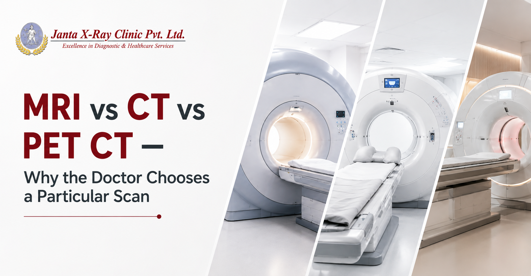
A prescription for an MRI, CT or PET CT can look deceptively simple. A few words on a referral slip, perhaps a body part and a particular protocol, can make it seem like a choice between three different machines. For the doctor, however, that recommendation usually represents a much longer chain of clinical reasoning. What could be causing the symptoms? Which structures could be involved? Is the concern about anatomy, soft tissue, an injury, blood vessels, disease activity or a change that needs to be followed over time?
The scan is chosen because of the question that needs to be answered. This is why two patients can walk into a consultation with a similar complaint and leave with completely different imaging referrals. One may be advised an MRI, another a CT, while a patient undergoing a particular cancer evaluation may be advised a PET CT. The difference is not necessarily the severity of the symptoms. It is often the information the clinician needs next.
At Janta X-Ray Clinic Pvt. Ltd., MRI, CT and PET CT form part of a wider diagnostic imaging service, with each examination serving different clinical requirements. The technology matters, but the reason for using it matters just as much.
MRI: High Resolution Imaging of Soft Tissues
MRI has a particular strength when the clinical question involves soft tissues and structures that require detailed visualisation. MRI uses a magnetic field and radio waves to produce detailed images and does not use ionizing radiation. It can be used to assess the brain, spine, joints, muscles, ligaments, tendons and several other soft tissue structures.
Consider someone with persistent knee symptoms. The question may not simply be whether the knee hurts. The clinician may need to assess cartilage, ligaments, tendons or other structures to investigate what could be contributing to the problem. The same principle applies to neurological symptoms. When the clinical question concerns the brain, spinal cord or particular soft tissues, MRI can provide the level of structural detail that may be needed.
This is why describing MRI simply as “the better scan” misses the point. Its value comes from the kind of information it can provide when that information is relevant to the clinical question. The symptom provides the reason for investigation, but the suspected cause helps determine what needs to be seen.
CT Scan: Precision of Anatomy and Quickness
Now consider a very different clinical situation. A patient arrives after significant trauma, and there may be concern about internal bleeding or injuries that cannot be assessed adequately from the outside. In such circumstances, time matters. CT can acquire detailed cross sectional images rapidly, making it particularly useful in many emergency and trauma situations.
Its role extends far beyond emergencies. CT is widely used for evaluating the lungs, bones, chest, abdomen and blood vessels, among other areas. There are also CT examinations in which contrast material is used to make particular blood vessels, organs or tissues more clearly visible.
When a clinician recommends a CT scan, the reasoning may therefore have as much to do with how quickly and precisely certain anatomical information is needed as with the body part being examined. CT is not simply an alternative to MRI. In particular clinical situations, its speed and the type of information it provides can make it the more appropriate examination.
PET CT Scan: Functional and Metabolic Assessment
Some clinical questions cannot be answered by looking at anatomy alone. This is where PET CT has a distinct role. The CT component shows anatomical structures and where a finding is located, while the PET component provides information related to metabolic activity. When these two forms of information are brought together, clinicians can assess both the location of a finding and its metabolic behaviour.
This becomes particularly relevant in many areas of cancer care. Depending on the clinical situation, PET CT may be recommended to assess the extent of disease, identify areas involved by cancer, evaluate response to treatment or investigate suspected recurrence. It also has selected applications in neurological and cardiac medicine.
The reason for recommending PET CT is therefore not simply that it is a more sophisticated CT scan. It is because the clinical team may need information about both where something is and how it is behaving metabolically. That additional information can be important when making decisions about the next stage of evaluation or treatment.
At Janta X-Ray Clinic Pvt. Ltd., PET CT scan is available for clinical situations where this combination of anatomical and metabolic information is relevant to the treating team. It is important to note that PET CT is not required for every person with cancer. The type of cancer, stage, treatment plan and clinical objective all influence whether the examination has a role.
One Symptom Can Lead to More Than One Possible Investigation
This is where the difference between medical imaging and an internet search becomes particularly important. Take a headache. The word itself tells a clinician very little. Where is the pain? How long has it been present? Is it new? Is it associated with visual symptoms, weakness, fever, trauma or other neurological findings? Is there a relevant medical history?
The answers can change the clinical thinking and, consequently, the imaging recommendation. The same is true of back pain, abdominal pain, breathing difficulties and many other common complaints. A symptom is a starting point for medical assessment, not an imaging prescription.
This is why searches such as “MRI or CT for headache” can be misleading when taken out of context. The question is not simply which scan can show a particular body part. The question is which examination can provide information relevant to the clinical concern. A search engine can describe what an MRI or CT does, but it cannot replace the clinical reasoning behind a referral.
Sometimes Different Scans Work Together
MRI, CT and PET CT are sometimes discussed as competing technologies, but clinical practice is not always an either or decision. Different examinations can provide different pieces of information at different stages of an investigation.
A CT may provide important anatomical information at one stage, while an MRI may subsequently be recommended because a particular structure needs closer assessment. In cancer care, CT or MRI may provide structural information while PET CT adds information about metabolic activity. These examinations can therefore complement one another rather than compete with one another.
The decision to use one examination or more than one is not about ordering more tests for the sake of it. It is about obtaining the information required at that point in the patient's care.
The Most Important Part of an Imaging Referral Is Not the Name of the Scan
MRI, CT and PET CT represent different capabilities within modern diagnostic imaging. MRI can provide detailed information about many soft tissues and internal structures. CT can deliver rapid and detailed cross sectional anatomical imaging. PET CT can combine anatomical information with information about metabolic activity.
But none of these descriptions, on their own, explains why a particular patient has been referred for a particular scan. That explanation lies in the clinical question. What does the doctor suspect? What needs to be confirmed or ruled out? What part of the body needs closer assessment? Is there a previous finding that needs comparison? Is the clinician assessing disease extent, treatment response or a new change?
These questions sit behind the referral.
At Janta X-Ray Clinic Pvt. Ltd., diagnostic imaging is therefore not simply about performing a scan. It is about providing imaging information relevant to the clinical requirement, supported by appropriate imaging protocols and reporting.
So the next time you see MRI, CT or PET CT written on an investigation referral, look beyond the name of the examination. There is a reason it was chosen, and that reason usually begins with a much more important question: What does the clinician need to find out?
FAQs
Question: Why can two doctors recommend different scans for similar symptoms?
Answer: Similar symptoms can have different possible causes. Doctors consider symptoms alongside examination findings, medical history, previous investigations and the conditions they are considering. Different clinical questions can therefore lead to different imaging referrals.
Question: Why might MRI be recommended when a CT has already been performed?
Answer: A CT may provide important information about anatomy, while MRI can provide additional detail about particular soft tissues or structures. A second examination may therefore be recommended because the clinician needs a different type of information.
Question: Why can PET CT be useful when CT has already shown a tumour?
Answer: CT can show the location and anatomical characteristics of a tumour. PET CT can add information about metabolic activity and may help in selected situations when clinicians need to assess disease elsewhere, treatment response or suspected recurrence.
Question: Can an imaging scan provide a diagnosis on its own?
Answer: Imaging findings are interpreted in the context of the patient's symptoms, clinical examination, medical history and other investigations. The radiology report contributes important information to the overall clinical assessment, but it is not considered in isolation.
Question: Why does a clinician sometimes ask for previous imaging?
Answer: A current scan can be more informative when previous images are available for comparison. Changes over time can provide important clinical information, particularly when monitoring a known finding or assessing treatment response.
Question: Does every cancer patient need a PET CT?
Answer: No. PET CT has specific clinical uses and is not automatically required for every cancer patient. The cancer type, stage, treatment plan and purpose of the examination influence whether PET CT is recommended.
Question: Why is CT often used in emergency situations?
Answer: CT can produce detailed cross sectional images quickly. In appropriate emergency situations, this speed can help clinicians assess injuries, internal bleeding and other urgent conditions.
Question: Is MRI always the first choice for soft tissue problems?
Answer: Not necessarily. MRI is particularly useful for many soft tissue examinations, but the appropriate imaging modality depends on the specific structure, suspected condition and clinical question.
If your clinician has recommended an MRI, CT or PET CT, Janta X-Ray Clinic Pvt. Ltd. provides diagnostic imaging services across a range of clinical requirements, with imaging examinations selected according to the investigation advised.


.jpg)
.jpg)
.jpg)
.jpg)


.jpg)
Comments List