
Incidental Findings on CT, MRI & Ultrasound: What They Mean
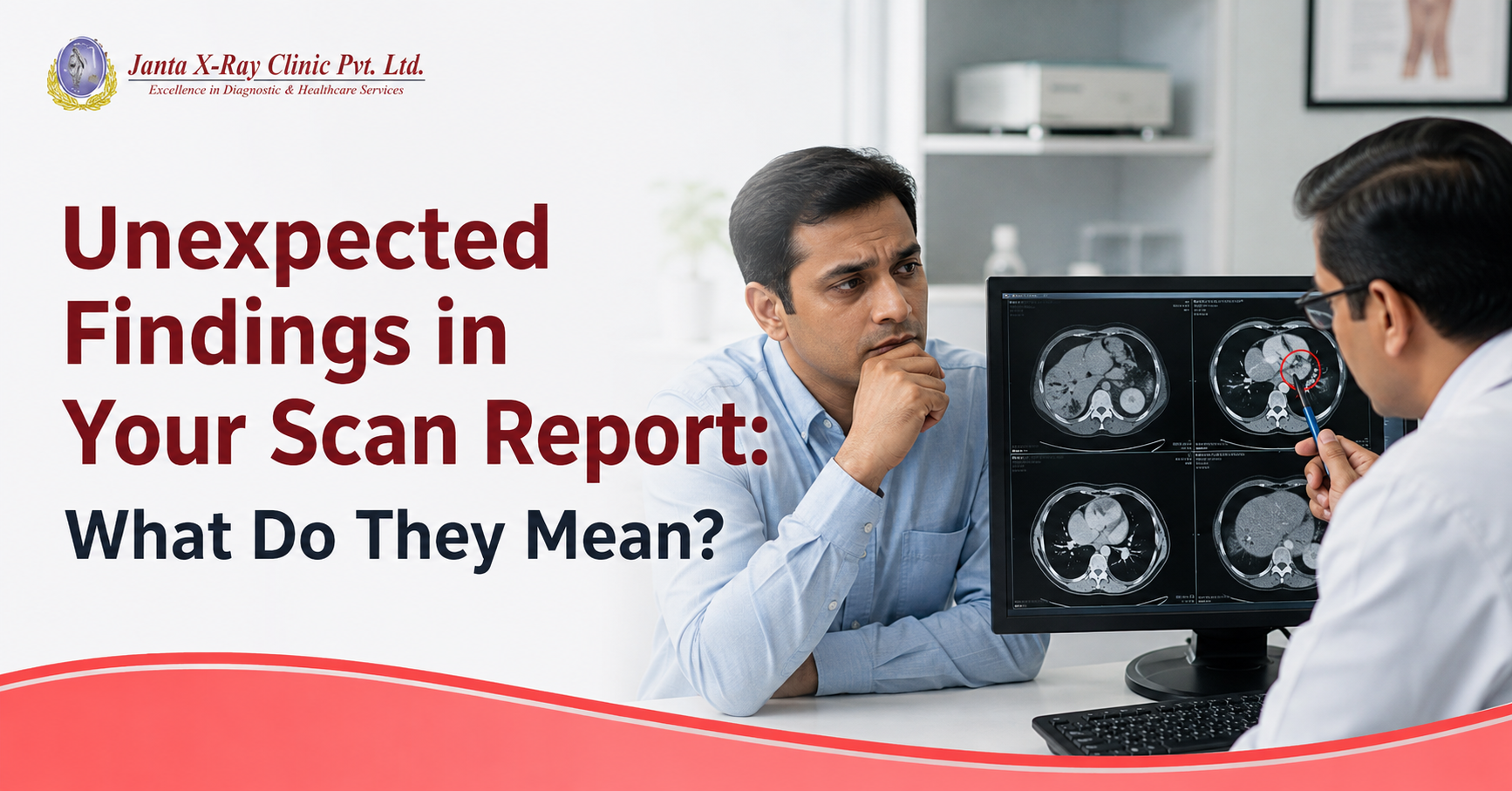
You went for a scan because of a specific problem. Maybe it was a headache, persistent stomach pain or back pain that would not go away.
You expected the report to explain that problem.
Instead, you notice words such as cyst, nodule, lesion or calcification mentioned in the report.
Naturally, the first thought is: “What does this mean?”
This is where incidental findings can cause unnecessary worry. An incidental finding is simply something noticed during an imaging examination that was not the original reason for the scan. It can happen during a CT scan, MRI or ultrasound, particularly when detailed imaging reveals something outside the main area of concern.
The important thing to remember is that an incidental finding is not automatically a disease or a diagnosis.
What is an incidental finding?
An incidental finding in radiology is an unexpected observation made while examining images for another clinical concern.
For example, a CT scan performed to investigate abdominal pain may also show a small kidney cyst. An MRI performed for back pain may reveal an unrelated finding. An ultrasound examination may identify a cyst or nodule that was not the reason for the examination.
Some incidental findings are common and harmless. Others may need to be monitored or investigated further.
So, the word “incidental” only explains how the finding was discovered. It does not tell you whether the finding is serious.
That distinction is important because a radiology report can contain medical terms that sound alarming without necessarily indicating a serious condition.
Why can a scan find something you were not looking for?
A CT, MRI or ultrasound examination is performed to answer a particular clinical question. But the images can show much more than the specific abnormality that the doctor was looking for.
Modern imaging provides detailed views of organs, tissues and other structures. While reviewing those images, a radiologist may notice an additional finding.
This is why incidental findings on CT scans, MRI scans and ultrasound examinations are not unusual.
Finding something unexpected does not necessarily mean that something has gone wrong. In some cases, the additional information may simply need to be documented. In others, it may help the doctor decide whether any further assessment is appropriate.
Does an incidental finding mean cancer?
No.
This is one of the most common concerns patients have after reading their scan report.
Words such as “lesion,” “nodule” or “mass” describe what is seen on imaging. They do not, on their own, confirm cancer.
For example, a renal cyst can be benign. Thyroid nodules can have different causes, and a pulmonary nodule may require assessment based on its size, appearance and the patient's individual risk factors.
The same word can also have very different significance depending on where it appears and what the images show.
That is why it is important not to interpret a single term from a radiology report without considering the rest of the report and your clinical history.
What happens when an incidental finding is reported?
There is no single approach for every incidental finding.
In some cases, the radiologist may consider the finding benign and not recommend any further investigation. In another situation, follow up imaging may be suggested to see whether the finding changes over time.
Sometimes, another imaging examination may provide more information. Your doctor may also recommend a laboratory test, clinical examination or consultation with a specialist, depending on the finding.
The decision can depend on several factors, including:
- The location of the finding
- Its size and appearance
- Your age
- Your symptoms
- Your medical history
- Your risk factors
- Whether it was seen on an earlier scan
This is why an incidental finding cannot be assessed properly from one word in a report.
Why your previous scans can make a difference
One of the most useful questions after an unexpected finding is:
“Was this present before?”
If an older CT, MRI or ultrasound is available, it may allow the radiologist or treating doctor to compare the finding with previous images.
A finding that has remained unchanged over time may be approached differently from one that is new or has changed.
So, if you have previous imaging reports or images, keep them available. They may provide useful information when your current scan is being assessed.
Should you worry if your report mentions a cyst, nodule or lesion?
Not necessarily.
These terms are used to describe different appearances seen on imaging.
A cyst may contain fluid and can be benign.
A nodule refers to a small, more defined area of tissue that appears different from the surrounding tissue.
A lesion is a broad term used to describe an area of abnormal or different appearance.
None of these words, by themselves, tell you what the final diagnosis is.
What matters is the complete imaging appearance and the clinical context in which the finding was discovered.
Why the radiologist's interpretation matters
Seeing an abnormality on an image is only part of diagnostic imaging.
A radiologist reviews the images, considers the reason for the examination and interprets what is seen. The final report brings those observations together and may indicate whether any further evaluation is appropriate.
This is particularly important when an examination produces a large number of detailed images.
At Janta X Ray, diagnostic imaging services include CT scans, MRI scans and ultrasound examinations, along with other imaging services. The centre provides a range of examinations across different clinical requirements, allowing patients to access diagnostic imaging under one roof.
If your doctor has recommended a CT, MRI or ultrasound, the purpose is not simply to produce images. The findings need to be interpreted in the context of your clinical concern so that your doctor can decide what to do next.
What should you do after reading an unexpected finding?
The first step is simple: don't jump to the worst conclusion.
Read the complete report rather than focusing on one unfamiliar word. Pay particular attention to the impression or conclusion section, where the radiologist summarises the important findings and recommendations.
Then discuss the report with the doctor who advised the examination.
If follow up has been recommended, ask what the recommendation means and when it should be done. If you have previous scans, share them with your doctor or imaging team for comparison when appropriate.
And if something in the report is unclear, it is completely reasonable to ask:
“What does this finding mean in my case?”
That question is far more useful than trying to diagnose yourself from an isolated search result.
The important thing to remember
An unexpected finding does not automatically mean an unexpected illness.
A CT scan, MRI or ultrasound can sometimes reveal something that was not causing your symptoms and was not the original reason for the examination. Some findings need no further action. Others may need monitoring or additional evaluation.
What matters is understanding what was found, how significant it is and whether anything needs to happen next.
If you have been advised to undergo a CT scan, MRI or ultrasound, Janta X Ray provides diagnostic imaging services for a range of clinical requirements. You can contact the centre to learn more about the appropriate examination recommended by your doctor.
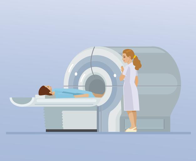
Janta X Ray offers MRI examinations including MRI brain, spine and contrast enhanced studies. Your doctor can advise which MRI examination is appropriate for your symptoms and clinical requirement.


.png)

.jpg)
1.png)

.jpg)
.jpg)
Comments List